
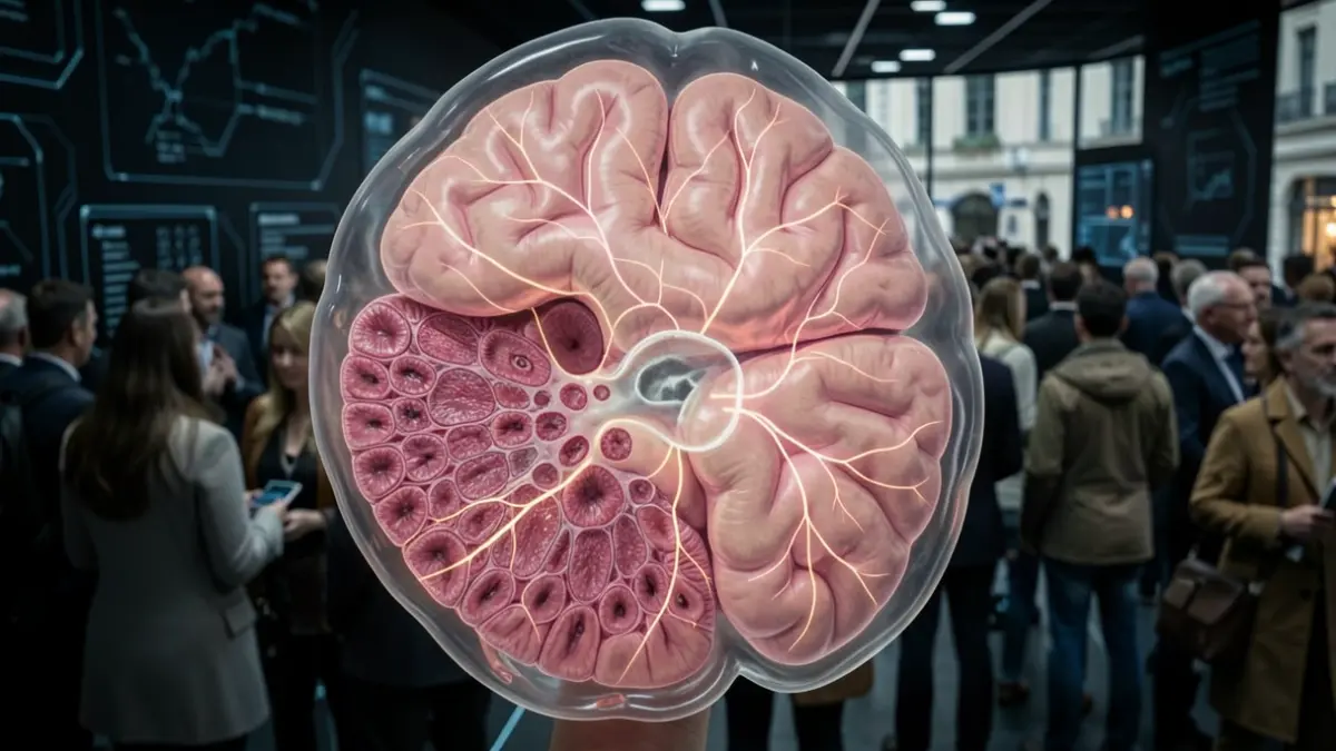
The study, published in Nature Neuroscience, reveals that sensory neurons and those related to motor and associative functions follow different developmental trajectories from early stages. Furthermore, researchers identify the Sp9 gene as an essential regulator for visual neurons to correctly acquire their identity.
The thalamus is a key brain structure that not only transmits information to the cerebral cortex but also integrates signals from the senses and participates in the organization of sensory maps. The Development, Plasticity, and Reprogramming of Sensory Circuits laboratory, led by researcher Guillermina López Bendito at the Institute of Neurosciences, located on the Sant Joan d’Alacant campus of the UMH, has contributed to revealing this active role of the thalamus over recent years.
This region is formed by different nuclei, with groups of neurons specialized in sensory, motor, and associative functions. However, questions remain about how they form during embryonic development and what determines the identity of their neurons. In this study, collaboration with researcher José López Atalaya, director of the Cellular Plasticity and Neuropathology laboratory, also integrated into the Institute of Neurosciences, has allowed the reconstruction of these cells' trajectories and the identification of the genetic programs involved in their differentiation.
To study how the different types of thalamic neurons are formed, the team analyzed individual cells from embryonic mouse brains at different developmental stages. This thalamus map has allowed them to follow the trajectory of thousands of neurons from their earliest stages to their final specialization.
The team was able to study each cell separately and determine which genes are active in it, as well as identify which regions of DNA are accessible for regulating those genes. “By integrating these two layers of information, we could reconstruct how cells differentiate over time and identify the genetic regulation programs that control these trajectories,” explains López Atalaya.
One of the main findings of the work is that thalamic neurons do not follow a single specialization route. The data reveal two major lineages that diverge very early during embryonic development: one will give rise to sensory nuclei and the other to nuclei related to motor and associative functions. Furthermore, both genetic programs progress at different rates, so that sensory circuits reach certain developmental stages before those associated with cognitive and motor functions.
Additionally, within the sensory pathway, the team discovered an unexpected intermediate stage: before specializing, some neurons go through a molecular state similar to those forming the higher-order nuclei of the thalamus. Unlike first-order nuclei, which transmit information from the senses, such as sight or touch, to the cerebral cortex, higher-order nuclei participate in more complex communication circuits between brain regions.
To confirm that the cells followed this trajectory, the researchers used a genetic marking technique that allows tracking the fate of progenitor cells and the neurons they generate. “This allowed us to experimentally validate the cell relationships we had predicted and discover that, within the sensory lineage, cells first pass through a state similar to higher-order nuclei before becoming sensory neurons, such as visual or somatosensory ones,” explains researcher Lorenzo Puche Aroca, the study's first author.
From the genetic programs identified in the atlas, the team pinpointed the Sp9 gene, which contains the information to produce the SP9 protein, a transcription factor that regulates the activity of other genes. It acts as an essential regulator for certain thalamic neurons to acquire a specific visual identity. To test its function, researchers experimentally reduced its activity during mouse embryonic development: “We wanted to go further and check if they really played a role in the specialization of their neurons. By reducing Sp9 activity, we saw that visual neurons did not correctly acquire their identity. This demonstrates that this gene plays a key role in establishing the characteristics that distinguish these neurons during development,” explains López Bendito.
The results of this study provide a new tool for studying how the thalamus is organized during development and how its circuits and functions are established. This knowledge offers a reference framework for better understanding certain alterations that may be related to neurodevelopmental disorders and neurological diseases.
This work was made possible thanks to funding from the Ministry of Science, Innovation and Universities and the State Research Agency; the European Research Council, under the Horizon 2020 program (ERC Advanced Grant SPONTSENSE); the "La Caixa" Foundation; the PROMETEO program of the Generalitat Valenciana; and the Severo Ochoa Program for Centers of Excellence.